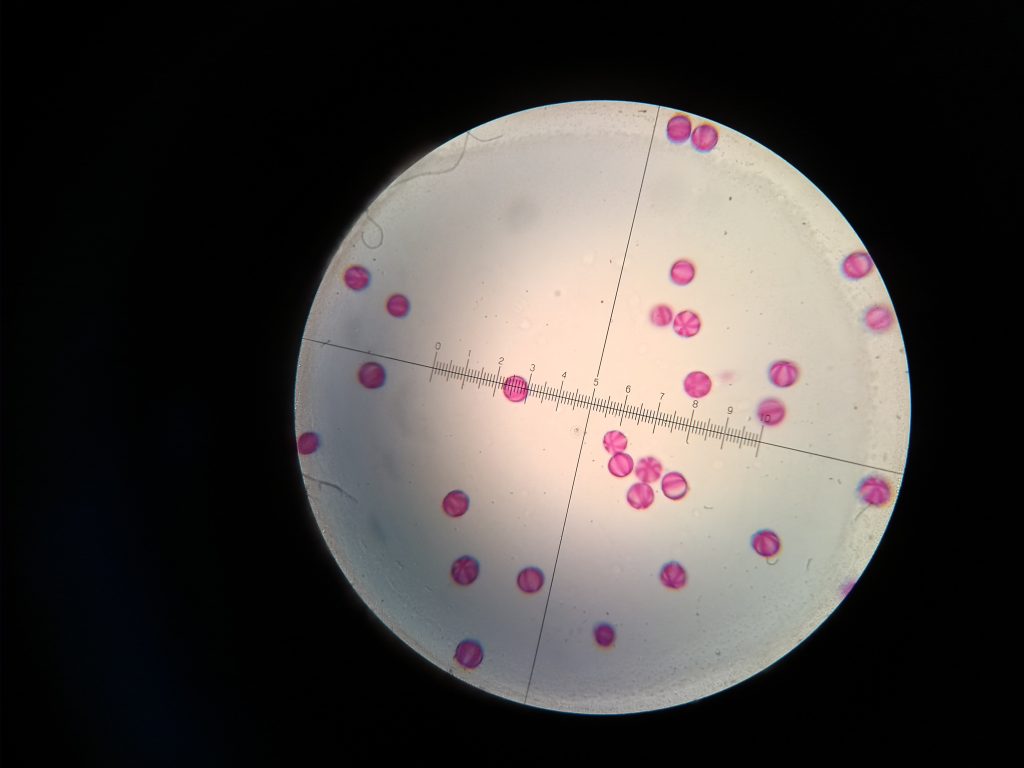
Photomicrograph of pollen grains (please provide details of what flower the pollen was collected from).
| Entrant ID: | 1042567 | Entrant Name: | Gill Brewer |
| Description: | Pollen grains from Phacelia tanacetifolia. X400 magnification. Photograph taken with a mobile phone through the eyepiece. A blue pollen load, dropped by a bee, was collected from the monitoring tray under a mesh floor. The pollen load was crushed with a drop of water. A sample was placed on a glass microscope slide and dried on a hotplate. Stain, glycerine jelly and coverslip were added, and the slide viewed under a light microscope. Pollen grains can by identified by their colour, size, shape, apertures and surface pattern. These pollen grains which looked like tiny pink sea-urchins, were confirmed as coming from Phacelia, as suspected from the blue colour of the pollen. |
||
| Entry: |  |
||
| Result: |  |
||
| Entrant ID: | 1044039 | Entrant Name: | John MacDougall |
| Description: | 1. This is a surface view of a pollen grain of Winter Jasmine (Jasminum nudiflorum) showing a medium-fine net surface and a long furrow with an aperture at the top. It is round and about 50 microns in diameter. Microscope: Brunel SP22, 400x magnification Camera: Motorola 3rd generation, hand-held 2. This is a pollen grain of French Marigold (Tagetes patula) showing a coarse net surface and many long, thin spines. It is round and about 38microns in diameter (medium size) Microscope: Brunel SP22, 400x magnification Camera: Motorola 3rd generation mobile phone, hand-held |
||
| Entry: |  |
||
| Result: |  |
||
